American Heart Center, Dubai Healthcare City Best Echocardiogram Test
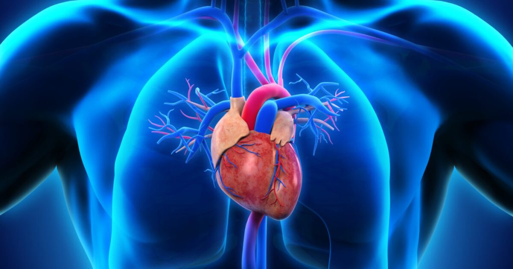
An echocardiogram is a test that uses sound waves to create detailed pictures of the heart. The images and information it provides are more comprehensive than a standard x-ray. Our best Echocardiogram test in Dubai delivers accurate results without exposing you to radiation.
American Board-Certified Cardiologists at American Heart Center Cardiology perform and interpret three common types of echocardiograms.
- A transthoracic echocardiogram (TTE) is non-invasive. A technician places a probe on the outside of the chest to create images of the heart. It uses color-flow Doppler ultrasound to create moving images of the heart. In certain situations, images obtained by this method are not detailed enough.
- A Doppler echocardiogram test in Dubai measures the speed and direction of blood flow. It provides a clearer view of how blood moves through the heart. Often used in combination with transthoracic methods, a Doppler echocardiogram reveals more detailed images than a basic echocardiogram.
- A stress echocardiogram uses ultrasound imaging to test how well your heart and blood vessels are working, before and during exercise. If a patient cannot perform light exercise, the doctor may give medication to stimulate the heart to pump harder.
Your doctor may suggest an echocardiogram to look at your heart’s structure and check how well it functions. If you experience CHEST PAIN, PAIN IN YOUR ARMS, HEART ATTACK, HIGH BLOOD PRESSURE, PALPITATIONS, NECK, BACK OR JAW PAIN, SHORTNESS OF BREATH, FATIGUE, WEAKNESS AND NUMBNESS, AN IRREGULAR HEARTBEAT OR SWOLLEN ARMS, LEGS, FEET, OR ANKLES are signs your doctor will look for when assessing you.
An echocardiogram may be done for further evaluation of signs or symptoms that may suggest:
- Heart Attack when an artery that sends blood and oxygen to the heart is blocked. Fatty, cholesterol-containing deposits build up over time, forming plaques in the heart’s arteries. If a plaque ruptures, a blood clot can form. The clot can block arteries, causing a heart attack.
- A gradual clogging of the arteries by fatty materials and other substances in the blood stream. It can lead to problems in the wall motion or pumping function of your heart.
- Cardiomyopathy, an enlargement of the heart caused by thick or weak heart muscle, diagnosed with the best Echocardiogram test in Dubai.
- Congenital heart disease. Defects in one or more heart structures that occur during formation of the fetus, such as a ventricular septal defect (hole in the wall between the 2 lower chambers of the heart).
- Heart failure. This condition weakens or stiffens the heart muscle during relaxation, preventing it from pumping blood efficiently. This can cause fluid buildup (congestion) in the blood vessels and lungs, and edema (swelling) in the feet, ankles, and other parts of the body.
- Aneurysm. A widening and weakening of a part of the heart muscle or the aorta (the large artery that carries oxygenated blood out of the heart to the rest of the body). The aneurysm may be at risk for rupture.
- Heart valve disease. Malfunction of one or more of the heart valves that may cause an abnormality of the blood flow within the heart. The valves can become narrowed and prevent blood from flowing through the heart or out to the lungs and body. The valves can also become leaky with blood flow leaking backwards. An echocardiogram can also check for infection of the heart valve tissue.
- Cardiac tumor. A tumor of the heart that may occur on the outside surface of the heart, within one or more chambers of the heart , or within the muscle tissue (myocardium) of the heart.
- An inflammation or infection of the sac that surrounds the heart.
Pericardial effusion or tamponade.
- The sac around the heart can become filled with fluid, blood, or infection. This can compress the heart muscle and prevent it from beating and pumping blood normally. This can cause symptoms of feeling dizzy, lightheaded, or a dangerous drop in blood pressure.
- Atrial or septal wall defects. Irregular channels between the right and left sides of the heart may be present at birth, or may occur from trauma, or after a heart attack. These defects occur in the upper filling chambers (atria) or the lower pumping chambers (ventricles). This may cause heart failure or poor blood flow, or increase your risk for stroke.
- Doctors can detect shunts in atrial and ventricular septal defects, as well as when irregular blood flow moves through the circulation from the lungs and liver.
The best echocardiogram test in Dubai may also be done to assess the heart’s overall function and general structure. Your doctor may have other reasons to recommend an echocardiogram. A normal echocardiogram reveals normal heart valves and chambers and normal heart wall movement
Have A Question On Your Mind ?
Our expert team is dedicated to providing answers and compassionate care for all your cardiac needs, ensuring you receive the best possible heart health guidance and support.

An abnormal echocardiogram can mean many things. Some abnormalities are very minor and do not pose major risks. Other abnormalities are signs of serious heart disease. You will need more tests by a specialist in this case. It is very important to talk about the results of your echocardiogram test with your provider, as we have team of the best cardiologist in Dubai.